
Mohs surgery is a specialized type of surgery utilized for the treatment of certain types of skin cancers. Considered a state of the art technique, it boasts the best rate of cure in complicated cases of basal cell and squamous cell carcinomas.
Advantages of Mohs surgery include two factors. The first advantage is that, with this procedure, the dermatologist only takes a small amount of normal cells, greatly increasing the probability of maintenance of skin function and reduction of scarring. The second advantage is that there are considerably less risks involved with using local anesthesia as compared to general anaesthesia.
History of Mohs Surgery
Mohs Surgery acquired its name from the founder and originator of the procedure, Dr. Frederic Mohs. While he was a young medical student, in the early 1930s, Mohs performed cancer research studies while working beside his medical instructor and mentor, Dr. Michael Guyer. Guyer was aware of a type of frozen tissue preparation for obtaining microscopic slides, and he also wrote a manual explaining a method of harvesting (processing) tissues for examination under a microscope. This book concentrated on the requirements of examination and documentation of these microscopic findings.
At a later time, Dr. Mohs used Guyer’s techniques to actually map out cancer cells surrounding blood vessels, nerves, bone and/or muscles. The tumors he observed were excised by shaving, or a process known as the saucerizing technique. This method involved removing the cancerous tumor, as a thin tissue disc, so that the affected area and the surrounding white blood cells could both be observed microscopically.
The Five Steps of Mohs Surgery
Most of the time, dermatologists use Mohs surgery as a treatment option to remove complicated instances of basal cell or squamous cell carcinomas. Occasionally, they use it for excising superficial cases of melanoma. There are five steps used in the Mohs surgical technique.
These include:
Step 1: Administration of Anesthetic
The dermatologist injects the skin cancer site using local anesthesia to totally numb the affected tissues. Use of a local anaesthetic has less inherent risks than general anesthetic.
Step 2: Removal of Any Visible Cancer Tissue
Any noticeable cancer tissue is carefully scraped with a semi-sharp medical instrument known as a curette. This device assists with the definition of margins separating tumor cells from healthy tissue. The doctor only removes the first thin layer of tissue. Steps may need to be taken to stop any bleeding.
Step 3: Tumor Map
After removing the first tissue layer, “tissue mapping” is performed as well as its orientation to other tissues and body parts. Tissue mapping acts as a guide to exact tumor coordinates. With the mapping process, affected tissues are labeled and coded according to color and correlated with their mapped positions. Sections of tissue are carefully examined to check for any cancer cells that may have been overlooked.
Step 4: Removing all Cancer Tissue
If necessary, after the analyzing the results of the tissue mapping, another thin layer is removed from the exact area where cancer was originally detected. The doctor does the same mapping and color coding process, and examines the tissue for remaining cancer cells. If even more cancer tissue is detected, the same procedures are repeated on subsequent tissue layers until all cancer is gone.
This precise technique allows for optimal preservation of as much normal tissue as possible. There is removal of the tissue which surrounds the cancerous area. Since Mohs surgery uncovers skin cancer roots, it has the best odds of completely removing the tumor while saving normal tissue(s). Cure rates with Mohs surgery are generally 99% for initial skin cancers and 95% for recurring tumors.
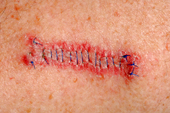
Step 5: Reconstructive Surgery
Following surgery, reconstructive techniques are personalized to maintain normal function while maximizing cosmetic results. The doctor can only determine the best method of wound repair after the cancer is totally removed. Occasionally, the wound will be allowed to heal on its own. Frequently, stitches are utilized to close the wound or a skin graft (flap) covers the area.
What to Expect with Mohs Surgery
Surgery is performed on an outpatient basis and usually takes about four hours to complete. It can take longer depending on the extent of the tumor(s). Preparation entails cleansing of the operative site, outlining it with a special pen, and an injection of local anaesthetic.
During the procedure, the doctor uses a scalpel to remove visible portions of cancer and also a thin layer of underlying tissue slightly larger than the tumor. The tissue is taken to the laboratory for precise microscopic analysis. This will take approximately one hour, and the patient goes to a waiting area.
If any tumor remains, surgery continues with another layer of tissue being removed. As much healthy skin tissue is left intact as possible. The entire process is repeated until tissue samples are free of cancer. An anesthetic agent will be repeated as needed. After the procedure, the surgeon and patient discuss the best reconstructive method for that individual.